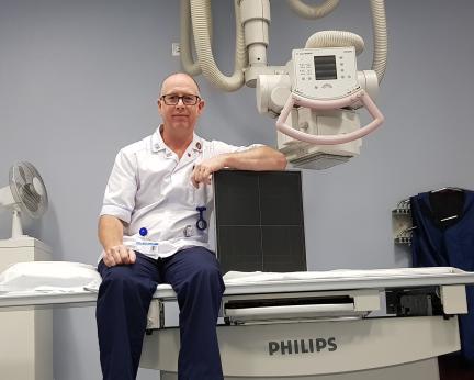
As a diagnostic radiographer I instantly make a difference to the outcome of someone’s treatment. Besides taking ‘plain film’ X-rays, I also spend time in operating theatres imaging for urology, orthopaedic and spinal surgeries in my day-to-day role.
I have chosen to further specialise in interventional radiography, performing real-time fluoroscopic (moving) imaging of gastro-intestinal and vascular procedures. Examples include patients who come into the theatre jaundiced and have stones removed, or straight from an ambulance in the middle of having a heart attack and leave with a fully-functioning heart!
Our real-time X-ray imaging gives the surgeons a view of what is happening inside the body. Because the soft tissue of the gastro-intestinal and vascular systems does not readily show up on X-ray, a radiopaque contrast dye is injected which shows up well on the X-ray, giving the surgeon a really detailed view of the area being operated on.
For instance, a blocked or narrowed cardiac artery will be instantly identifiable by the omission of contrast dye, and when the artery has been reopened, this is confirmed by the completeness of the artery when further dye is injected. This imaging allows the surgeon to operate through a small incision, usually in the groin, rather than opening up the chest cavity, and the patient will be up and walking again within a few hours.


