Imaging (non-ionising)
Non-ionising imaging is an area of healthcare science that includes ultrasound, magnetic resonance imaging (MRI) and optical imaging.
As a clinical scientist in this area, you’ll develop new procedures, provide safety advice, perform quality assurance activities, teach and train, develop image analysis and reconstruction software, and also undertake more general research and development activities.
Overview
You will typically be based within either a radiology or medical physics department of a hospital.
Non-ionising imaging techniques are generally safe which is especially reassuring when imaging sensitive patient groups, such as children.
The non-ionising imaging theme also encompasses some treatment modalities which are supported by clinical scientists, including ultra violet treatments for skin conditions and laser surgery. You’ll play an important role in ensuring these treatments are delivered safely, help to develop ways of improving them or introduce new treatments.
Working life
If you work in non-ionising imaging, you’ll use a range of imaging techniques, including:
- ultrasound - uses high frequency soundwaves that can produce real-time images of tissues and organs within the body. Ultrasound is commonly used to monitor fetal development and to assess and screen patients who present with a variety of clinical indications including suspected blood flow problems, gall stones and tumours
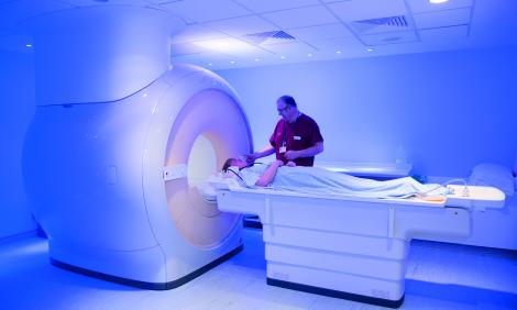
- MRI - uses a combination of strong magnetic fields and radiowaves. MRI is renowned for its ability to differentiate soft tissues. It can be used to examine almost any part of the body and is the modality of choice for imaging the central nervous system. MRI has the ability to characterise normal and abnormal tissue and can be used to produce a wide variety of different contrasts which may reflect blood flow, cellular density, tissue stiffness through to changes in blood oxygenation, which has been extensively used to understand brain function
- optical imaging - involves measuring the physical properties of light to help make a diagnosis. For example, when shining particular wavelengths of light onto cancerous tissues, they may exhibit different absorption, light scattering or fluorescence behaviours to normal skin. This is an area which is continuing to move from the laboratory into the clinic. Optical systems are currently being evaluated to assess brain function and breast cancer. There are also a number of dedicated optical systems which are used to evaluate eye disease
Who will I work with?
You will be part of a team that includes radiologists, diagnostic radiographers and sonographers.
Want to learn more?
-
Most jobs in the NHS are covered by the Agenda for Change (AfC) pay scales. This pay system covers all staff except doctors, dentists and the most senior managers. As a clinical scientist working in non-ionising imaging, your salary will be between AfC bands 6 and 9, depending on your role and level of responsibility. Trainee clinical scientists train at band 6 level, and qualified clinical scientists are generally appointed at band 7. With experience and further qualifications, you could apply for posts up to band 9.
Staff will usually work a standard 37.5 hours per week. They may work a shift pattern. Terms and conditions of service can vary for employers outside the NHS.
-
With further training or experience or both, you may be able to develop your career further and apply for vacancies in areas such as further specialisation, management, research, or teaching.
Non-ionising techniques are extensively used in research, for example, to investigate the effectiveness of new drugs.
-
Job market
In November 2018, there were 6,123 clinical scientists registered with the Health and Care Professions Council.
The NHS Scientist Training Programme (STP) attracts many more applicants than there are places and so there is considerable competition for places.
Finding and applying for jobs
When you’re looking for job vacancies, there are a number of sources you can use, depending on the type of work you’re seeking.
Check vacancies carefully to be sure you can meet the requirements of the person specification before applying and to find out what the application process is. You may need to apply online or send a CV for example.
For the STP there is an annual recruitment cycle. Applications usually open in early January for the intake in the following autumn and should be made through the National School of Healthcare Science's website, where you can also find information about the programmes and the recruitment process.
Key sources relevant to vacancies in the health sector:
- vacancies in organisations delivering NHS healthcare can be found on the NHS Jobs website
- opportunities in the Civil Service can be found on the Civil Service Jobs website
As well as these sources, you may find suitable vacancies in the health sector by contacting local employers directly, searching in local newspapers and by using the Universal Jobmatch tool.
Find out more about applications and interviews.
Volunteering is an excellent way of gaining experience (especially if you don’t have enough for a specific paid job you’re interested in) and also of seeing whether you’re suited to a particular type of work. It’s also a great way to boost your confidence and you can give something back to the community.
-
For further information aboit working and training in non-ionising imaging, please contact:


